달서구 치과 추천, 임플란트 후 잇몸이 내려가는 4가지 이유
Why Gums May Recede Around Dental Implants: Four Things to Check
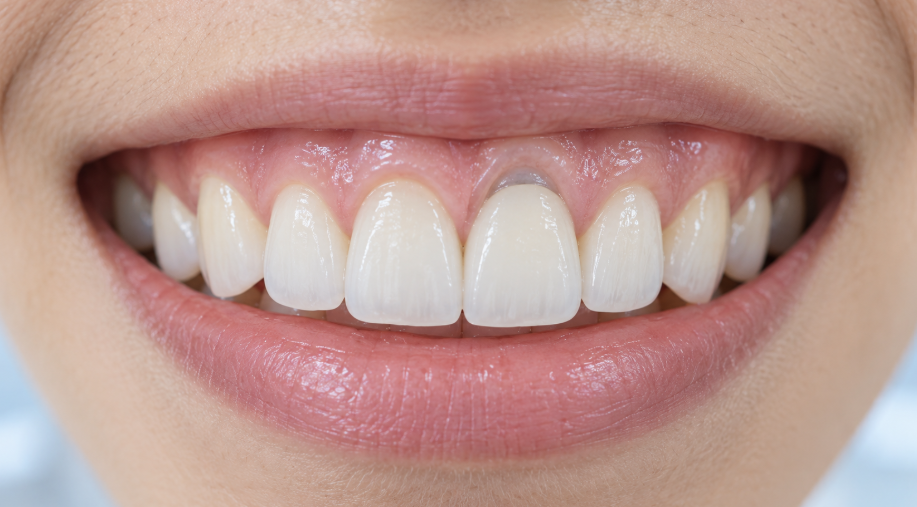
임플란트 주변 잇몸이 내려가 보이거나 금속색이 비칠 때, 확인해야 할 4가지 원인과 검진·치료 방향을 정리했습니다.
This article explains four factors to assess when gum recession or a gray metallic hue is noticed around a dental implant, along with possible examination and management directions.
안녕하세요. 서울바른플란트치과 대표원장 문홍열입니다.
Hello. I am Dr. Hong-Yeol Moon, the chief director at Seoul Bareun Plant Dental Clinic.
임플란트 치료 후 시간이 지나면서 잇몸선이 아래로 내려가 보이거나, 잇몸 가장자리에서 회색빛이 비쳐 걱정되어 내원하는 분들이 있습니다.
특히 앞니는 작은 변화도 눈에 잘 띄기 때문에 기능에는 큰 불편이 없어도 심미적으로 민감하게 느껴질 수 있습니다.
Some patients visit because, over time after implant treatment, the gumline appears to have moved downward or a gray hue becomes visible near the gum margin. Small changes are particularly noticeable in the front teeth, so they may be cosmetically concerning even when there is little functional discomfort.
먼저 말씀드리면, 임플란트 주변 잇몸이 내려갔다고 해서 곧바로 임플란트가 실패했거나 제거가 필요한 것은 아닙니다.
다만 잇몸과 뼈의 두께, 임플란트의 3차원 위치, 염증 및 골소실 여부, 보철물의 형태와 청소 환경을 나누어 확인해야 치료 방향을 합리적으로 정할 수 있습니다.
To begin with, gum recession around an implant does not automatically mean that the implant has failed or must be removed. A sound treatment plan requires an assessment of soft- and hard-tissue thickness, the implant's three-dimensional position, inflammation or bone loss, and the restoration's shape and cleansability.
임플란트 주변 잇몸이 내려가는 현상은 무엇인가요?
What Does Gum Recession Around an Implant Mean?
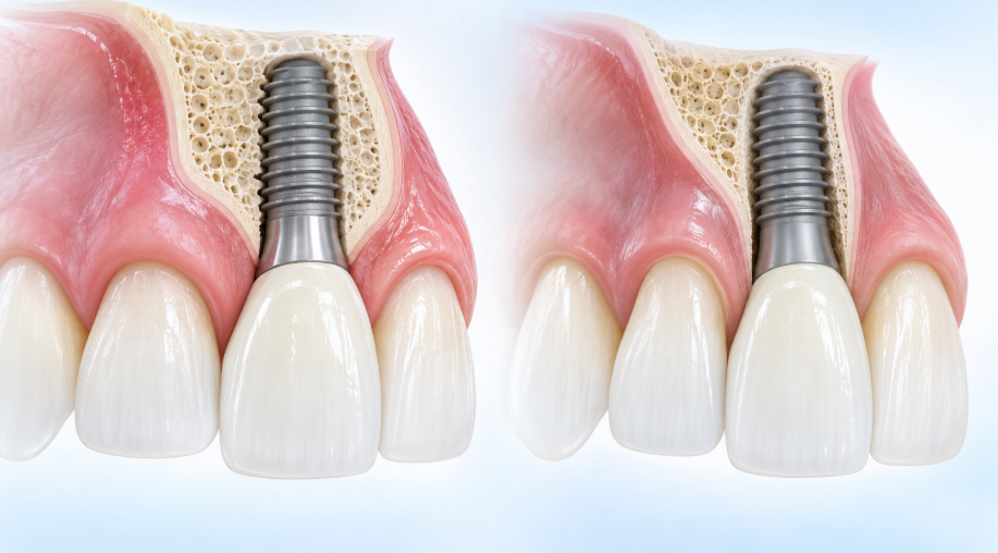
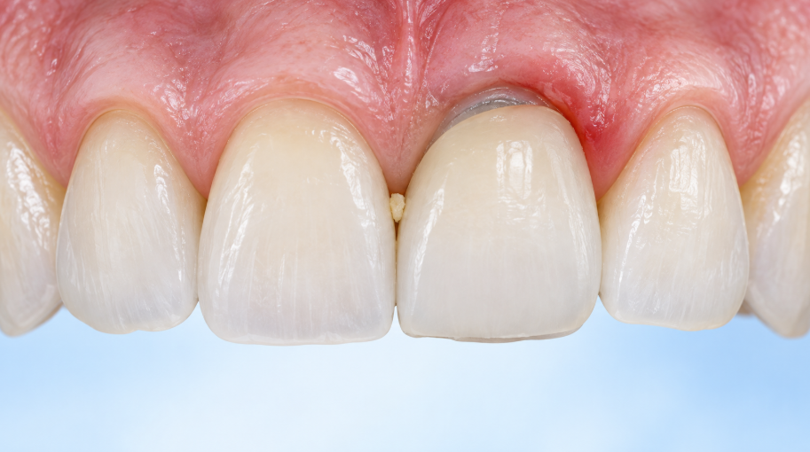
임플란트 주변의 잇몸 가장자리가 원래보다 아래쪽으로 이동해 크라운이 길어 보이거나, 지대주·임플란트의 회색빛이 드러나는 현상을 임상적으로 임플란트 주변 연조직 퇴축 또는 연조직 결손으로 설명할 수 있습니다.
When the gum margin around an implant moves downward so that the crown appears longer or the gray color of the abutment or implant becomes visible, it may be clinically described as peri-implant soft-tissue recession or a soft-tissue deficiency.
자연치의 잇몸 퇴축과 비슷해 보이지만, 임플란트는 주변 조직의 두께와 뼈의 지지, 식립 위치, 보철물 형태에 따라 양상이 달라질 수 있습니다. 따라서 거울로 보이는 변화만으로 원인을 단정하기보다, 과거 사진·방사선 사진과 비교해 실제로 진행 중인지부터 확인하는 과정이 중요합니다.
Although it can resemble gum recession around a natural tooth, the pattern around an implant may differ depending on tissue thickness, bony support, implant position, and restoration design. Rather than deciding the cause from appearance alone, it is important to compare prior photographs and radiographs to determine whether the change is actually
먼저, 이렇게 구분합니다.
A Practical First Check
보이는 변화 또는 증상우선 확인할 내용판단의 방향
| 잇몸선만 살짝 내려가 보임 | 잇몸 두께, 바깥쪽 뼈의 지지, 이전 사진과의 변화 | 조직 형태·위치 문제 여부 평가 |
| 금속색 또는 지대주가 비쳐 보임 | 연조직 두께, 보철물 경계와 색조 | 심미적 문제와 안정성 함께 확인 |
| 출혈·붓기·고름·냄새가 동반됨 | 잇몸주머니, 플라크, 방사선상 골소실 | 점막염·주위염 가능성 평가 |
| 음식물이 자주 끼고 닦기 어려움 | 크라운의 볼륨, 인접면·치간 공간, 교합 | 보철물 형태와 관리 환경 점검 |
What you may noticeWhat to check firstDirection of assessment
| The gumline alone looks slightly lower | Gum thickness, support from the outer bone, and change from earlier photographs | Assess tissue form and implant-position factors |
| A gray metallic color or abutment is visible | Soft-tissue thickness, restoration margin, and color | Assess cosmetic concerns together with tissue stability |
| Bleeding, swelling, pus, or odor is present | Peri-implant probing, plaque accumulation, and radiographic bone change | Assess for possible peri-implant mucositis or peri-implantitis |
| Food frequently becomes trapped and cleaning is difficult | Crown contour, contact areas and interdental space, and occlusion | Review restoration design and the hygiene environment |
1. 잇몸과 뼈가 원래 얇은 경우입니다.
1. The Gum and Outer Bone May Be Naturally Thin
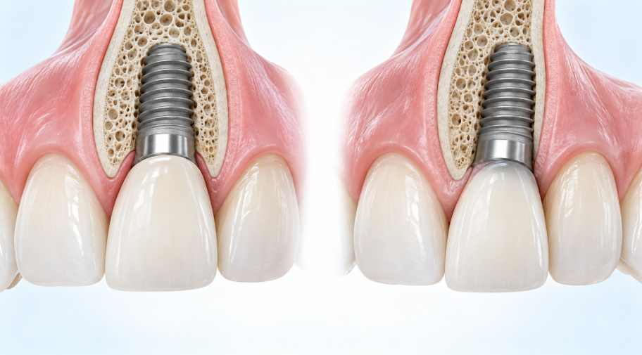
임플란트 바깥쪽의 뼈와 잇몸이 얇은 경우에는 시간이 지나면서 잇몸선 변화가 더 잘 보일 수 있습니다.
잇몸 조직이 얇으면 실제 이동량이 크지 않아도 지대주나 임플란트의 회색빛이 비쳐 심미적으로 더 크게 느껴지기도 합니다.
When the outer (buccal) bone and gum tissue around an implant are thin, a change in the gumline may be easier to notice over time. With thin soft tissue, even a small change can allow a gray hue from the abutment or implant to show through and appear more pronounced cosmetically.
특히 앞니 부위는 바깥쪽 뼈가 얇은 경우가 적지 않아, 잇몸선의 높이뿐 아니라 잇몸의 두께와 색조, 인접 치아와의 조화를 함께 살펴야 합니다.
발치와 동시에 식립한 경우에도 원래 조직의 두께와 치유 양상에 따라 변화가 나타날 수 있어 경과 관찰이 필요할 수 있습니다.
The outer bone in the front-tooth area is often thin. For this reason, evaluation should include not only the level of the gumline but also tissue thickness, color, and harmony with neighboring teeth. Even when an implant was placed at the time of extraction, the original tissue thickness and healing pattern can affect later changes, so follow-up observation may be needed.
얇은 조직 자체가 곧 병을 뜻하는 것은 아닙니다.
염증이나 진행성 골소실 없이 안정적으로 유지된다면, 심미적 요구와 관리 가능성을 고려해 유지 관찰할 수 있습니다.
Thin tissue itself does not necessarily indicate disease. If the area is stable without inflammation or progressive bone loss, monitoring may be appropriate after considering cosmetic expectations and the ability to maintain hygiene.
2. 임플란트가 바깥쪽으로 위치했거나 기울어진 경우입니다.
2. The Implant May Be Positioned Too Far Toward the Outside or at an Unfavorable Angle
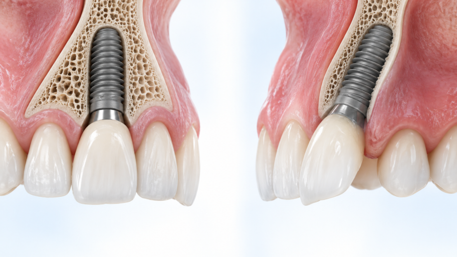
임플란트가 치조골의 바깥쪽에 너무 가깝게 위치하거나 각도가 불리하면, 바깥쪽 뼈와 잇몸이 충분한 지지를 받기 어려울 수 있습니다.
이 경우 시간이 지나며 잇몸선이 내려가 보이거나 금속색이 노출될 가능성을 검토하게 됩니다.
If an implant is positioned too close to the outer side of the jawbone or placed at an unfavorable angle, the outer bone and gum may not have sufficient support. In such cases, the gumline may appear to recede over time or a gray metallic color may become visible.
여기서 중요한 점은 잇몸만 보고 판단하지 않는 것입니다.
임플란트의 위치, 남아 있는 뼈의 형태, 크라운이 나오는 방향과 부피를 함께 확인해야 합니다.
잇몸을 덮는 처치를 먼저 고려하더라도, 뼈와 임플란트 위치의 지지가 충분하지 않다면 장기적인 안정성을 따로 검토해야 할 수 있습니다.
The key point is not to assess the gum alone. The implant position, shape of the remaining bone, and the direction and volume of the emerging crown should be reviewed together. Even if soft-tissue coverage is considered first, long-term stability may need separate evaluation when bony support or implant position is insufficient.
식립 위치가 원인으로 의심된다고 해서 무조건 제거를 결정하지는 않습니다.
증상, 조직 두께, 골소실 진행 여부, 보철물의 위치, 환자분의 심미적 요구를 종합해 보철 수정·연조직 처치·재수술 여부를 개별적으로 판단합니다.
Suspected positioning issues do not automatically lead to implant removal. The need for restoration modification, soft-tissue treatment, or reoperation is considered individually by integrating symptoms, tissue thickness, whether bone loss is progressing, restoration position, and the patient's cosmetic concerns.
https://booking.kakao.com/detail/ticket/726439?t_src=sharing
서울바른플란트치과의원
서울바른플란트치과의원 진료예약
booking.kakao.com
3. 임플란트 주변 염증과 골소실이 동반된 경우입니다.
3. Inflammation and Bone Loss May Be Present Around the Implant
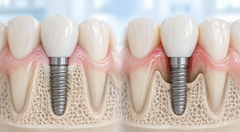
잇몸이 내려가면서 출혈, 붓기, 고름, 냄새, 반복되는 불편감이 함께 나타나거나 방사선 사진에서 뼈 변화가 관찰된다면 임플란트 주위 조직의 염증 여부를 확인해야 합니다.
임플란트 주위염은 염증과 함께 지지 뼈가 진행성으로 소실되는 상태이므로, 단순히 ‘잇몸이 덮일 수 있는가’보다 염증과 세균막을 먼저 조절하는 것이 우선입니다.
If gum recession occurs with bleeding, swelling, pus, odor, repeated discomfort, or radiographic bone changes, inflammation in the tissues around the implant should be assessed.
Peri-implantitis is an inflammatory condition accompanied by progressive loss of supporting bone, so controlling inflammation and bacterial biofilm takes priority over simply trying to cover the recessed gum.
다만 수술 후 초기 치유 과정에서 보일 수 있는 골 변화만으로 곧바로 주위염이라고 단정할 수는 없습니다.
보철물이 완성된 뒤의 기준 방사선 사진과 잇몸주머니 검사 결과를 비교해, 변화가 진행하는지와 염증 신호가 있는지를 함께 보아야 합니다.
However, bone changes seen during early healing after surgery should not by themselves be diagnosed as peri-implantitis. A baseline radiograph taken after the final restoration is completed and the results of peri-implant probing should be compared to determine whether change is progressing and whether inflammatory signs are present.
출혈만으로 주위염을 확정할 수는 없지만, 부드러운 검사에서 반복적으로 출혈이 보이거나 고름·붓기·깊어진 잇몸주머니가 동반되면 검진을 미루지 않는 것이 좋습니다. 통증이 없다고 해서 항상 안정적이라고 판단할 수 있는 것도 아닙니다.
Bleeding alone cannot confirm peri-implantitis. However, when bleeding recurs during gentle probing or is accompanied by pus, swelling, or deeper peri-implant pockets, it is best not to delay an examination. The absence of pain does not always indicate that the area is stable.
4. 보철물 형태와 청소 환경도 함께 살펴야 합니다.
4. Restoration Shape and the Cleaning Environment Also Matter
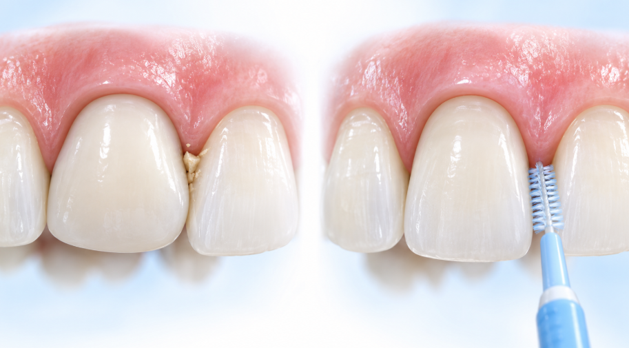
크라운이 잇몸 쪽에서 지나치게 볼록하거나 음식물이 머무르기 쉬운 형태라면 칫솔·치실·치간 관리가 어려워질 수 있습니다.
관리가 어려운 환경은 세균막이 쌓이기 쉬운 조건이 될 수 있으므로, 잇몸이 내려간 부위에서는 임플란트 자체뿐 아니라 보철물의 윤곽과 인접면, 치간 공간까지 함께 점검해야 합니다.
If a crown is overly bulky near the gumline or shaped in a way that traps food, brushing, flossing, and interdental cleaning can become difficult. A difficult-to-clean environment can promote biofilm accumulation, so evaluation of a recessed area should include not only the implant itself but also the restoration contour, contact areas, and interdental spaces.
강한 이갈이·이악물기나 불균형한 씹는 힘이 의심되는 경우에는 보철물의 교합도 확인합니다.
힘 문제 하나만으로 잇몸 퇴축의 원인을 단정할 수는 없지만, 주변 조직과 보철물에 가해지는 부담을 줄이기 위한 조정이 필요할 수 있습니다.
When strong grinding, clenching, or uneven bite forces are suspected, the restoration's occlusion should also be checked. Force alone cannot determine the cause of gum recession, but adjustments may be needed to reduce the load placed on surrounding tissues and the restoration.
필요에 따라 보철물의 형태를 조정하거나 임시 보철물로 조직 형태를 유도한 뒤, 연조직 이식과 같은 치료를 함께 검토하기도 합니다.
Depending on the situation, the restoration contour may be adjusted or an interim restoration may be used to guide tissue shape before procedures such as soft-tissue grafting are considered.
달서구 치과에서 잇몸 퇴축을 확인할 때 보는 항목
What Is Checked When Evaluating Peri-Implant Gum Recession?
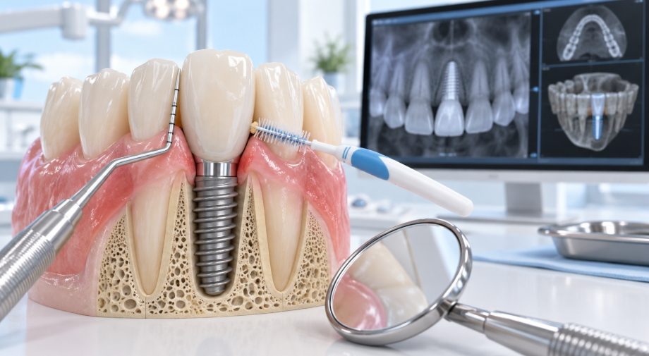
임플란트 주변 잇몸이 내려가 보인다면 다음 항목을 함께 확인하는 것이 도움이 됩니다.
When gum recession is noticed around an implant, it is helpful to assess the following points together.
잇몸선의 위치와 두께, 금속색 비침 여부
The location and thickness of the gumline, and whether a gray metallic hue is visible.
출혈·붓기·고름·냄새 등 염증 신호
Inflammatory signs such as bleeding, swelling, pus, or odor.
잇몸주머니 깊이와 이전 검사 결과의 변화
Peri-implant pocket depth and changes from earlier examination results.
방사선 사진에서 보이는 뼈 높이와 진행성 골소실 여부
Bone level on radiographs and whether bone loss is progressing.
필요 시 CT에서 확인하는 바깥쪽 뼈와 임플란트의 3차원 위치
When needed, the outer bone and three-dimensional implant position on CT.
크라운의 형태, 음식물 끼임, 교합과 청소 접근성
Crown contour, food impaction, occlusion, and access for cleaning.
CT는 모든 경우에 일률적으로 촬영하는 검사가 아니라, 일반 방사선 사진과 임상 검사를 바탕으로 위치·뼈 결손을 더 자세히 확인할 필요가 있을 때 활용할 수 있습니다.
CT is not routinely required in every case. It can be used when clinical examination and conventional radiographs indicate that implant position or a bone defect needs more detailed evaluation.
https://booking.kakao.com/detail/ticket/726439?t_src=sharing
서울바른플란트치과의원
서울바른플란트치과의원 진료예약
booking.kakao.com
잇몸이 내려갔다면, 치료는 어떻게 달라질까요?
How Can Management Differ When the Gumline Has Receded?
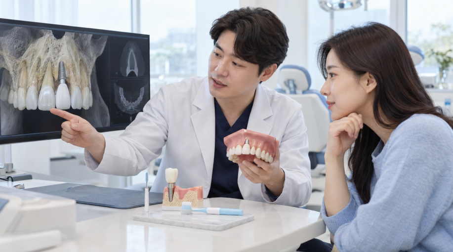
치료는 한 가지 방식으로 정해지지 않습니다.
There is no single treatment approach for every case.
- 염증과 골소실이 없고 변화가 안정적이라면 정기 검진과 위생관리, 사진 비교를 통해 경과를 관찰할 수 있습니다.
If there is no inflammation or bone loss and the change is stable, the area may be monitored with regular examinations, hygiene care, and comparison photographs.
- 플라크와 염증이 확인되면 전문적인 세정과 관리 방법 교육을 먼저 시행하고, 위험요인을 함께 조절합니다.
If plaque and inflammation are identified, professional cleaning and oral-hygiene instruction are addressed first, together with relevant risk factors.
- 보철물의 형태가 청소를 방해하거나 잇몸을 압박하는 경우에는 보철물 조정 또는 교체를 검토할 수 있습니다.
If the restoration shape interferes with cleaning or compresses the gum, modification or replacement of the restoration may be considered.
- 심미적 요구가 크거나 조직 두께가 부족한 경우에는 연조직 이식 등으로 두께와 윤곽을 보완하는 방법을 논의할 수 있습니다.
If cosmetic concerns are substantial or tissue thickness is insufficient, options such as soft-tissue grafting may be discussed to improve thickness and contour.
- 임플란트 위치나 뼈 결손이 치료 결과에 큰 영향을 주는 경우에는 추가 수술 또는 재치료의 필요성을 신중히 평가합니다.
When implant position or a bone defect is expected to substantially affect the outcome, the need for additional surgery or retreatment is evaluated carefully.
즉, 치료의 목표는 단순히 잇몸을 위로 올리는 데만 있지 않습니다.
염증을 조절하고, 뼈와 잇몸의 지지 상태를 확인하며, 장기적으로 관리하기 쉬운 보철 환경을 만드는 것이 함께 중요합니다.
In other words, treatment is not solely about moving the gumline upward. It is equally important to control inflammation, confirm the support provided by bone and gum tissue, and create a restoration environment that is easier to maintain over the long term.
이런 신호가 있다면 확인을 권합니다.
Signs That Merit an Examination
이전보다 잇몸선이 빠르게 내려가 보이는 경우
The gumline appears to be receding more rapidly than before.
금속색이 새롭게 보이거나 노출 범위가 넓어지는 경우
A gray metallic color has newly become visible or the exposed area is expanding.
칫솔질·치실 사용 시 출혈이 반복되는 경우
Bleeding recurs during brushing or flossing.
붓기, 고름, 냄새, 자발통, 음식물 끼임이 지속되는 경우
Swelling, pus, odor, spontaneous pain, or food impaction persists.
크라운이 흔들리거나 씹는 느낌이 달라진 경우
The crown feels loose or the bite feels different.
자주 묻는 질문
Frequently Asked Questions

Q. 임플란트의 금속색이 보이면 바로 제거해야 하나요?
Q. Does a Visible Gray Metallic Color Mean the Implant Must Be Removed Right Away?
그렇지 않습니다. 금속색 비침 또는 일부 노출만으로 제거 여부를 결정하지는 않습니다.
염증과 골소실의 유무, 임플란트 위치, 노출 정도, 보철물 상태, 환자분의 불편과 심미적 요구를 함께 평가해야 합니다.
No. A gray metallic hue or partial exposure alone does not determine whether removal is needed. Inflammation, bone loss, implant position, the extent of exposure, restoration condition, discomfort, and cosmetic concerns should all be assessed together.
Q. 잇몸에서 피가 나면 임플란트 주위염인가요?
Q. Does Bleeding From the Gum Mean Peri-Implantitis?
출혈은 확인이 필요한 신호이지만, 그것만으로 주위염을 확정할 수는 없습니다.
잇몸주머니 검사와 방사선 사진, 세균막 관리 상태를 함께 보아 점막염인지 주위염인지, 다른 자극 요인은 없는지 판단합니다.
Bleeding is a sign that warrants assessment, but it cannot confirm peri-implantitis by itself. Peri-implant probing, radiographs, biofilm control, and other possible irritants are considered together to distinguish peri-implant mucositis from peri-implantitis.
Q. 잇몸이 조금 내려갔는데 재수술이 꼭 필요한가요?
Q. Is Reoperation Always Necessary for Mild Gum Recession?
아닙니다.
변화가 안정적이고 염증이나 진행성 골소실이 없다면 유지 관찰과 보철물·위생관리만으로 관리할 수 있는 경우도 있습니다.
반대로 변화가 진행하거나 심미적·기능적 불편이 크다면 연조직 처치나 보철 치료를 검토할 수 있습니다.
No. If the change is stable and there is no inflammation or progressive bone loss, some cases can be managed with monitoring and restoration and hygiene care alone. In contrast, if the change progresses or creates substantial cosmetic or functional discomfort, soft-tissue or restorative treatment may be considered.
핵심 정리
Key Takeaways
임플란트 후 잇몸이 내려가는 현상은 단순 노화 한 가지로 설명하기 어렵습니다.
잇몸과 뼈의 두께, 임플란트 위치, 염증 및 뼈 변화, 보철물의 형태와 청소 환경이 복합적으로 영향을 줄 수 있습니다.
Gum recession around an implant is difficult to explain as aging alone. Gum and bone thickness, implant position, inflammation and bone changes, restoration design, and the cleaning environment can all play a role.
대구 치과 또는 달서구 치과를 알아보실 때도 ‘잇몸이 내려갔다’는 모습만으로 바로 재수술을 결정하기보다, 먼저 염증과 골소실이 있는지, 임플란트와 보철물의 위치는 적절한지, 현재 상태가 진행 중인지를 평가받는 것이 중요합니다.
When looking for dental care in Daegu or Dalseo-gu, it is important not to decide on reoperation solely because the gumline appears lower. First, have inflammation and bone loss, the position of the implant and restoration, and whether the condition is progressing assessed.
서울바른플란트치과에서는 임플란트 주변의 잇몸·뼈·보철물·교합 상태를 함께 확인한 뒤, 구강 상태에 맞는 관리 또는 치료 방향을 안내하고 있습니다.
At Seoul Bareun Plant Dental Clinic, we assess the gum tissue, bone, restoration, and occlusion around the implant together, then explain management or treatment directions appropriate to the individual's oral condition.
이 글은 일반적인 치과 건강 정보입니다. 실제 진단과 치료 방법, 치료 기간 및 예후는 임플란트 위치, 남아 있는 뼈와 잇몸의 상태, 염증 정도, 전신 건강, 위생관리 습관 등에 따라 달라질 수 있으므로 의료진의 진료와 상담이 필요합니다.
This article is for general dental-health information only. Diagnosis, treatment methods, treatment duration, and prognosis can vary according to implant position, remaining bone and gum condition, degree of inflammation, general health, and oral-hygiene habits. An in-person examination and consultation with a dental professional arery. necessary.
https://booking.kakao.com/detail/ticket/726439?t_src=sharing
서울바른플란트치과의원
서울바른플란트치과의원 진료예약
booking.kakao.com
'서울바른플란트치과 임상' 카테고리의 다른 글
| 임플란트 씹을 때 아픈 4가지 이유 (0) | 2026.10.21 |
|---|---|
| 달서구 치과, 치실과 치간칫솔 차이 (0) | 2026.10.20 |
| 대구 레진 치료 후 통증, 정상일까요? (0) | 2026.10.16 |
| 대구치과, 임플란트 보철물이 빠졌다면? (0) | 2026.10.15 |
| 대구치과, 정확한 양치법, 알고 계신가요? (0) | 2026.10.14 |


